For appointment and information, you can rich us on +90532 433 6003(whatsapp)
 Obstetrician, Gynecologist, Genital Esthetic Surgeon in Turkey
For appointment and information, you can rich us on +90532 433 6003(whatsapp)
 Obstetrician, Gynecologist, Genital Esthetic Surgeon in Turkey
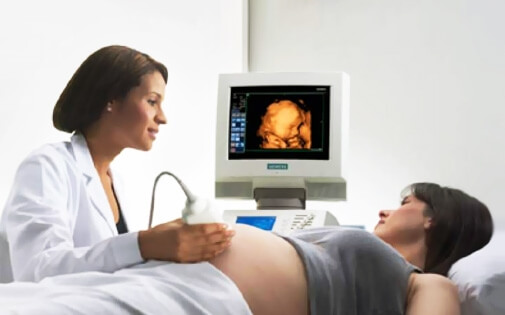
3D ultrasonography shows still pictures of the baby, whereas 4D ultrasonography reveals the baby live so that the parents can watch fetal movements such as kicking, thumb sucking, swallowing and even fascial expressions.

Ultrasound technology has developed so much rapidly in the recent years that we are almost able to see which parent the baby will take after in utero.
3D ultrasonography shows still pictures of the baby, whereas 4D ultrasonography reveals the baby live so that the parents can watch fetal movements such as kicking, thumb sucking, swallowing and even fascial expressions.
For parents watching their baby by ultrasound technology throughout pregnancy is an amazing feeling. However the main reason to use ultrasound technology should be to diagnose congenital birth defects,not entertainment.
Actually for the diagnosis of birth defects, 2D Ultrasonography is sufficient for most cases.2D ultrasound uses black and white pictures of the baby which show internal organs in different cross sections.
3D and 4D ultrasound is different from 2D Ultrasound, because the pictures are formed to show fetal skin rather than fetal organs. Therefore it is easy to recognize the baby’s facial expressions, yawning, thumb-sucking and tongue-sticking in appropriate fetal positions.
Ultrasonoghraphy devices use sound waves that are sent to the baby through an ultrasound probe and reflected back from the baby through the same probe, to be converted by a special software into ultrasound pictures. 4D technology uses many 2D pictures taken at different angles combined as a single image.
Current studies have shown that all three types of ultrasound technology (2D, 3D and 4D) are safe for the fetus when used for medical purposes.
There is a principle we use while performing ultrasound called ALARA (as low as reasonably achievable) This means we set our ultrasound machines so that we use the lowest amount of energy possible in the shortest duration necessary for the examination. Although there are no known hazardous effects of ultrasound technology, it is wise to expose the baby to the least amount of energy possible to avoid any possible long term side effects.
For ethical reasons, ultrasound technology should only be used by adequately trained professionals. In USA there are some facilities where 3D and 4D keepsake ultrasound devices are abused just for recording baby pictures and videos which is not recommended by the ISUOG (International Society of Ultrasound in Obstetrics and Gynecology) and ACOG (American College of Obstetricians and Gynecologists). FDA (Food and Drug Administration) also warns parents against such use of +D ultrasound technology.
It is important to keep in mind that ultrasound is not just for entertainment but for diagnosis. Even if you are interested in having your baby’s 4D ultrasound pictures and videos taken as a bonding experience or memento, make sure the examination is performed by a trained medical professionaL, because there is a possibility that an existing congenital malformation may be missed by the sonographer or a normal physiological finding may be misinterpreted as abnormal leading to parental anxiety.
As long as the baby’s position is suitable, it is possible to obtain 3D pictures or 4D videos at almost any gestational age. However if there is only one chance you can get a 4 D ultrasound examination, you can prefer to have it around 26-30 weeks of gestation.
In Turkey fetal 4D Ultrasound examinations can be performed either by medical doctors who have received residency training in Radiology or Obstetrics and Gynecology specialties. There is another subspecialty of Obstetrics and Gynecology named as Perinatology (Maternal Fetal Medicine) which gives extra advanced training in fetal ultrasonography examinations.
Perinatal medicine aims to detect problems as early as possible in utero so that they can either be treated or when there is a lethal congenital malformation, termination of pregnancy can be offered to the family as early as possible during the course of pregnancy.
A targeted ultrasound scan is a systemic examination of the fetus by a trained professional to be able to detect any congenital morphological malformations and genetic conditions. 2 D ultrasound examination is sufficient for a targeted ultrasound scan for most of the cases. Targeted ultrasound scan is performed at around 18-22 weeks of gestation.
Targeted ultrasound scan assesses fetal development and location.It detects fetal anomalies and when there is a lethal anomaly, termination of pregnancy may be discussed with the family as an option. For lethal congenital anomalies, medically indicated termination of pregnancy is a legal option in Turkey even after 10 weeks of gestation as long as there is a medical report signed by 3 doctors.
It is possible to see the internal organs, fetal skeleton and the outer surface and fetal skin on a 4D ultrasound scan.Most people think 4D ultrasound is designed to see only the fetal skin or face. However it is possible to visualise fetal organs by this technology for diagnostic purposes. This software can even be used for fetal echocardiography, which is a detailed examination of the fetal heart.
Four D ultrasound technology is available at Dr. Burcu Saygan Karamürsel Clinic who has been primarily trained as an Obstetrician and Gynecologist and additionaly as a Perinatologist (Maternal Fetal Medicine Specialist). She is a member of ISUOG (International Society of Ultrasound in Obstetrics and Gynecology) and ACOG (American Collegeof Obstetricians and Gynecologists) receiving continuous training in recent advances in ultrasound technology. She is also a professor at Atılım University Faculty of Medicine teaching.
If you are interested in having a fetal 4D ultrasound examination or you have been referred by your obstetrician for a targeted ultrasound scan, you can reach our clinic for an appointment through the following telephone numbers: +90312 219 2233, Whatsapp: +90545 219 2234